how do they x ray babies hips
The babys legs have differences in their lengths or appearances. Ive used tongue depressors and sponges to hold down squirming fingers and.

Pin Page
3 notes 2 X-ray of hip dysplasia is generally the initial imaging of choice thereafter.

. In the direct projection or frontal obtained by focusing the x-ray tube perpendicular to the body plane - front or rear and axial transverse or horizontal plane fixing the elements of the joint from top to bottom - along the femur. Soft tissues take in less radiation so they appear different. Parents are usually afraid to hold them as tight as necessary.
An X-ray technician will take pictures of the hip. Youll be asked to partly undress your baby and take off their diaper for the test. It s sometimes called congenital dislocation of the hip or hip dysplasia.
Its a cast that goes around both hips and down the leg to keep the hips aligned. If there is a tumour it shows up as an irregular. A hip X-ray is a safe and painless test that uses a small amount of radiation to make images of the hip joints where the legs attach to the pelvis.
No cuts are needed. Hip ultrasounds are a safe non-invasive procedure that does not use any radiation. If she does have it they may try to brace it first.
Most premature babies go on to. Around 6 months of age enough bone is present in an infant hip to make an X-ray more accurate than ultrasound. Your baby will be placed on a table on their back or side.
A hip click can be felt by the examiner when the hip joints may not have formed normally. Radiographers appear to like it. For a simple baby hand xray the child can usually be scanned using a standard device using just a light restraint.
It is put on by an orthopedic surgeon while using x. For a chest x-ray however babies and younger children need a special machine to ensure the radiation dose hits the right spot. It might help to feed your baby just before the ultrasound to make your little one more relaxed.
Those children would have gone untreated and perhaps would have developed early hip arthritis if they had not returned for a final visit. You will go in the room with him he will need to be stripped from the waist down they will take x-rays of him flat on his back legs dead straight and together you wil be able to hold him in this position then an x-ray of his still on his back with his knees bent facing outwards and the soles of his feet put together he will be fine its. After about 6 months of age x-rays are done because the bones are often too well developed to use ultrasound successfully.
Calcium in your bones takes in more radiation so your bones appear white on the X-ray. Without restraint they would inevitably wriggle around and blur the images. The doctor hears or feels a hip click when moving the infants thigh outward during a routine checkup.
You will go in the room with him he will need to be stripped from the waist down they will take x-rays of him flat on his back legs dead straight and together you wil be able to hold him in this. Two tests are performed called the Barlow and Ortolani tests to examine the function of the hip joints. Hip ultrasounds are recommended for infants from birth to six months old because their bones have not fully developed.
A hip X-ray radiograph is a medical imaging test that creates a picture of your hip joints and pelvic bones. Surprisingly approximately one in four had developed dysplasia that was treated with bracing. Instead of releasing them from care the children were re-examined with x-rays at four to six months of age.
Your How do they x ray babies uk images are available. Answer 1 of 5. If it persists they may put on a spica cast.
We wrap them and hold them or preferably their parents hold them. An X-ray of the pelvis focuses specifically on the area between your hips that holds many of your reproductive and digestive organs. Babies are flexible and very resilient.
During the examination an X-ray machine sends a beam of radiation through the pelvic bones and hip joints and an image is recorded on a computer or special film. If I suspect DDH in a baby when should I refer to a. Ultrasonography may be appropriate notes 2 6 months.
The maximum visual information is given by the x-ray of the hip joint in two projections. Ultrasonography of hip dysplasia is generally the investigation of choice at up to 4 months due to limited ossification of the skeleton. How do they x ray babies uk are a topic that is being searched for and liked by netizens today.
Ultrasounds are the diagnostic method of choice for infants under 6 months of age. The picture shows the inner structure anatomy of your hips in black and white.

Diapering And The Harness Cloth

How Parents Can Help Their Baby S Hip Dysplasia

Pin On Hip Dysplasia Pao

Congenital Hip Dislocation Chd Happens When A Child Is Born With An Unstable Hip Read On To Learn Dislocacion De Cadera Huesos De La Cadera Batido De Papaya

Anatomy Pathology Medicine Nursing Radiography Radiologictechnologist Radiology Radiologystudent I Radiology Student Medical Anatomy Radiology Schools

Radiology Radiologic Imaging Signs List Collection Illustrated Cases Xray X Ray Gi Gu Chest Thoracic Teaching Website Web Site

12 Incredible X Rays Reveal How Different Pregnant Animals Look
Pin Page

Xray Image Child Swallowed Coins Medical Stock Photo 342401210 Shutterstock

Posterior Hip Dislocation Radiology Case Radiopaedia Org

What Causes Hip Dysplasia International Hip Dysplasia Institute

Pin Page

Severe Hip Dysplasia In A Boxer The Red Arrows Are Pointing To The Over Growth Of Bone At The Femoral Neck Head The Red Arrow Shades Of Grey Hip Dysplasia

Pin On درمانی
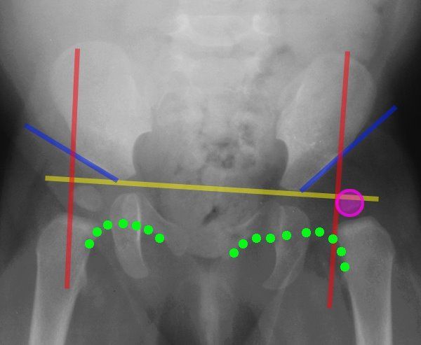
Lines Of The Hip Pediatrics

Pin Page

Pin On Adult Hip Dysplasia Awareness

Generalized Osteopenia Radiology Reference Article Radiopaedia Org

X Rays